XB-IMG-116835
Xenbase Image ID: 116835
|
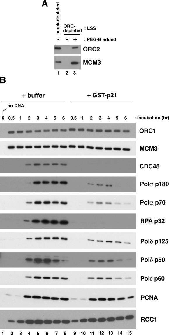
Figure 3. Analysis of the proteins bound to plasmid beads during incubation in Xenopus egg extracts. (A) pBluescript-coupled beads were incubated for 30 min in the mock-depleted (lane 1), the ORC-depleted extracts (lanes 2) or the ORC-depleted extracts supplemented with PEG-B fraction (lane 3). (B) pG5λ6.6-coupled beads or the beads alone (no DNA) were incubated in LSS for the indicated time periods in the absence or presence of 13 μg/ml GST-p21. After incubation, the proteins bound to the beads were analyzed by western blotting with the appropriate antibodies as indicated. Image published in: Zembutsu A and Waga S (2006) © 2006 The Author(s). Creative Commons Attribution-NonCommercial license Larger Image Printer Friendly View |