XB-IMG-125345
Xenbase Image ID: 125345
|
|
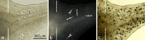
Fig. 8. Dopa staining in the 5-day regenerating tail in the mutant (amputated at stage 49). a,b The mutant regenerating tail before dopa staining observed under transmitted light (a), or incident light (b). c The mutant regenerating tail after dopa staining observed under transmitted light. Dashed lines indicate the amputation level. White pigment cells were appearing in the mutant regenerating tail on day 5 post-amputation (b). Dopa staining was observed in white pigment cells (arrowheads) as well as melanophore precursors which were not visible under transmitted light or incident light before dopa reaction (arrows) Image published in: Fukuzawa T (2010) © The Author(s) 2010. Creative Commons Attribution-NonCommercial license Larger Image Printer Friendly View |