XB-IMG-125838
Xenbase Image ID: 125838
|
|
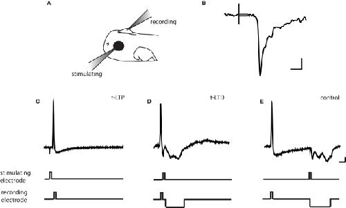
Figure 1. Protocols for inducing STDP. (A) Schematic of in vivo recording set-up. (B) Representative tectal response to retinal stimulation. Scale bar is 5pA and 5 ms. Black line indicates beginning of 100-ms stimulus and gray box indicates stimulus artifact. (C) t-LTP protocol. A 2 ms 0.17–0.2 pA current pulse was injected in the post-synaptic tectal cell to evoke an action potential 5–10 ms following the onset of the EPSC. (D) t-LTD protocol. The stimulating electrode in the retina was timed to evoke an EPSC 5–10 ms after an action potential in the tectal neuron. (E) In the control conditioning protocol, this timing window was lengthened to 100 ms, outside the described window for plasticity at this synapse (Zhang et al., 1998). A 20–60 ms hyperpolarizing current pulse was timed to prevent action potential firing due to the retinal EPSC. Traces are from cells held at −40 to −50 mV in current clamp. Scale bar is 10 mV and 20 ms. Image published in: Tsui J et al. (2010) Image downloaded from an Open Access article in PubMed Central. Copyright © 2010 Tsui, Schwartz and Ruthazer. Larger Image Printer Friendly View |