XB-IMG-123652
Xenbase Image ID: 123652
|
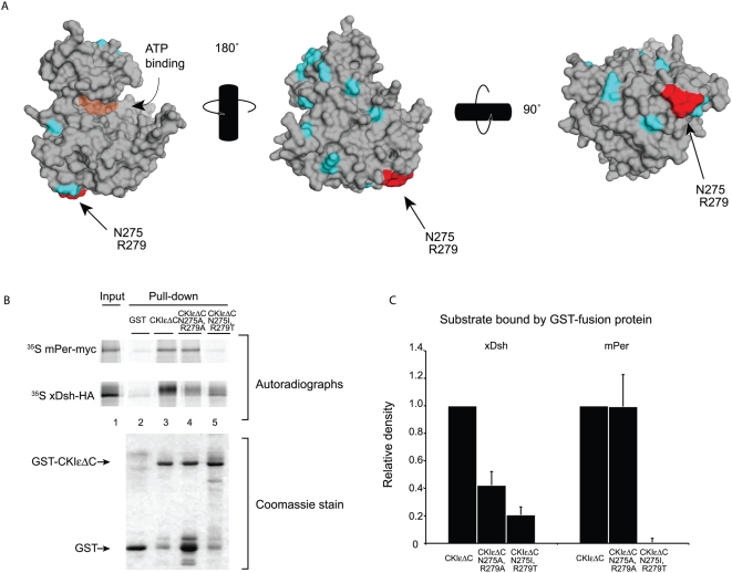
Figure 4. Residues 275 and 279 regulate binding to xDsh and mPer1.(A) Space-filling representation of CKIδ (PDB ID 1CKJ, [50]). Residues shown in cyan and red are conserved between CKIε and CKIδ, but not CKIα. Red residues N275 and R279 are solvent accessible and are chemically distinct in CKIα. Orange shading shows the position of the ATP binding cleft. (B) Binding of 35S-labeled mPer1 and xDsh to GST-CKIεΔC, GST-CKIεΔC N275A/R279A, or GST-CKIεΔC N275I/R279T was performed. (C) Quantification of three independent experiments. Values are normalized against the amount of protein bound by GST-CKIεΔC. Image published in: Dahlberg CL et al. (2009) Dahlberg et al. Creative Commons Attribution license Larger Image Printer Friendly View |