XB-IMG-169389
Xenbase Image ID: 169389

|
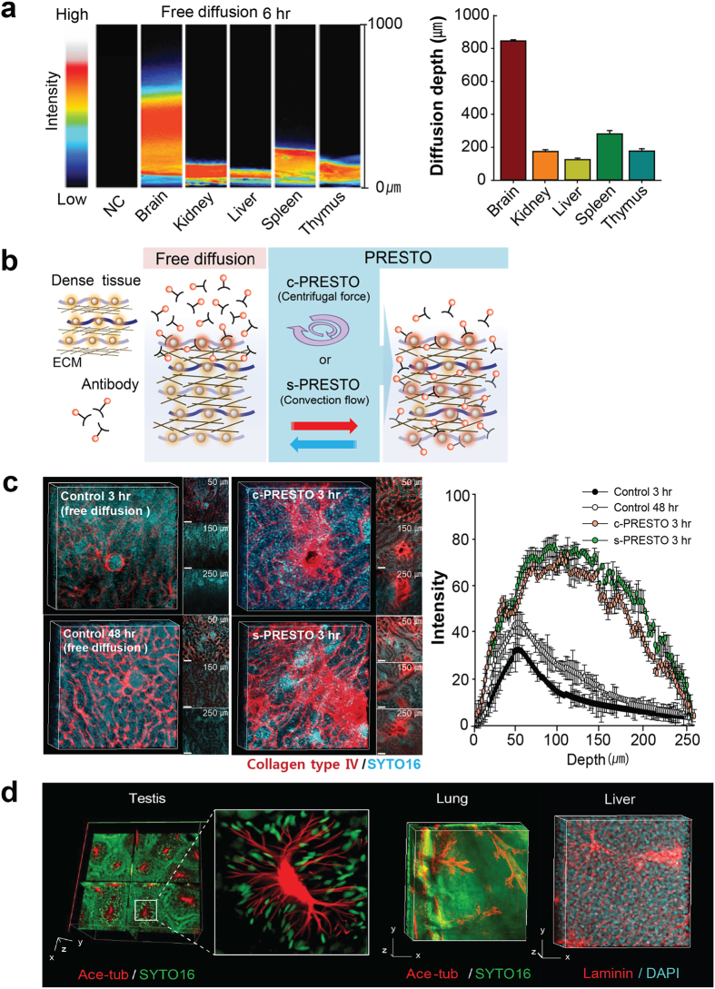
Figure 5. ACT-PRESTO (active clarity technique-pressure related efficient and stable transfer of macromolecules into organs) for rapid immunolabeling of dense tissues.(a) Comparison of diffusion rate using ACT-processed organs. (b) Schematic diagram for dense tissue immunohistochemistry. Tissues for centrifugal PRESTO (c-PRESTO) were centrifuged at 600 × g for 3 hours using a standard table-top centrifuge to expedite penetration of the primary and secondary antibodies. A syringe pump was used for the antibody reaction during syringe PRESTO (s-PRESTO). (c) Kidneys were labeled with collagen type IV using various protocols. Note that 3 hours of c- or s-PRESTO markedly enhanced the depth of specific labeling compared to that of the controls. Three-dimensional (3D) reconstructed images were obtained with a Zeiss 700 confocal microscope with a Plan-apochromat 10 ×/0.45 M27 lens, 2 × confocal zoom (stack size, 200 μm; stack step, 2 μm), and post-processed with Vaa3D software. Scale bar, 100 μm. Depth of fluorescence intensity was greater in PRESTO-treated tissue compared to that of free-diffusion labeled samples using ACT processed kidney tissue (mean ± standard deviation, n = 5). (d) Reconstituted 3D images of testis, lung, and liver. The organs were stained with acetylated tubulin (red in testis and lung) or laminin antibodies (red in liver). SYTO16 or DAPI were used for nuclear staining of the organs. Images were obtained with a Zeiss 700 confocal microscope with a Plan-apochromat 10 ×/0.45 M27 lens, 2 × confocal zoom (testis; stack size, 600 μm; stack step, 5 μm; liver; stack size, 226 μm; stack step, 2 μm; Scale bar, 100 μm), with a EC Plan-Neoflua 5 ×/0.16 M27 lens (lung; stack size, 1,265 μm; stack step, 5 μm; Scale bar, 500 μm). Larger Image Printer Friendly View |
