XB-IMG-178517
Xenbase Image ID: 178517
|
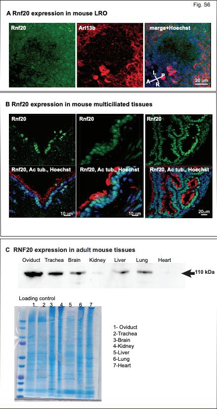
Figure S6: (A) Rnf20 expression in the mouse LRO. The LRO structure was identified by presence of monociliated cells labeled with anti-Arl13b, nuclei were visualized by Hoechst staining. Anterior (A)-Posterior (P) and Left (L)-Right (R) axes are indicated. The image is the maximum intensity projection of a Z stack encompassing the entire LRO.
(B) RNF20 expression in mouse multiciliated tissues visualized by immunostaining with anti-RNF20 antibody (Green). Multiciliated cells were identified by ciliary labeling with anti-acetylated tubulin (Red), and nuclei by Hoechst staining.
(C) Western blot of 20µg/lane adult mouse tissue probed with anti-Rnf20 antibody
showing expected 110kD band. Loading was normalized to total protein determined by BCA assay and confirmed by SimplyBlue SafeStain (Coomassie G-250). Image published in: Robson A et al. (2019) Copyright © 2019. Image reproduced with permission of the Publisher and the copyright holder. This is an Open Access article distributed under the terms of the Creative Commons Attribution License. Larger Image Printer Friendly View |