XB-IMG-124110
Xenbase Image ID: 124110
|
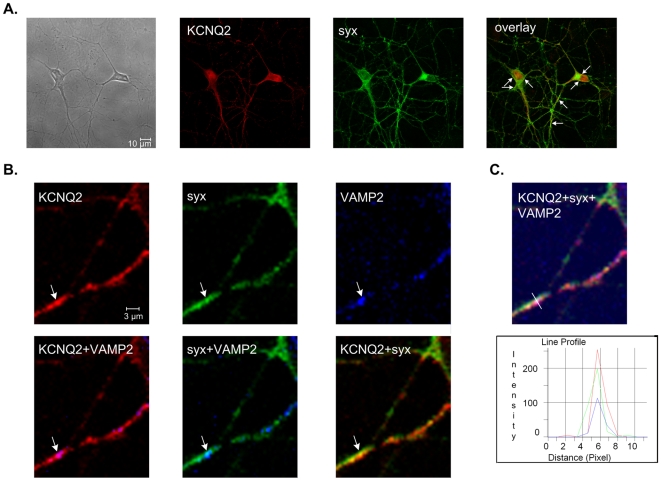
Figure 5. Colocalization of KCNQ2 and syntaxin 1A at synaptic sites marked by VAMP-2 immunoreactivity in hippocampal neurons.A, Immunocytochemistry experiments show colocalization (overlay, yellow) of KCNQ2 (red) and syntaxin 1A (syx; green) in rat hippocampal neurons. High colocalization areas of KCNQ2 and syntaxin 1A are indicated by arrows. B, Colocalization of KCNQ2, syntaxin 1A and VAMP-2 in rat hippocampal neurons as detected by triple immunocytochemistry and illustrated by the merge images. KCNQ2 (red), syntaxin 1A (green) and VAMP-2 (blue) are indicated in the top images from left to right. The bottom images from left to right show the colocalization of KCNQ2 and VAMP-2 (merge, pink), syntaxin 1A and VAMP-2 (merge, light blue) and KCNQ2 and syntaxin 1A (merge, yellow). A varicosity colocalized with VAMP-2, syntaxin 1A and KCNQ2 is indicated by arrow. C, The same image as in B showing all three markers; KCNQ2 (red), syntaxin 1A (green) and VAMP-2 (blue). A linescan was placed through the varicosity indicated by arrow in B. The varicosity was shown to colocalize all three signals and thus, is indeed a synaptic one. Image published in: Regev N et al. (2009) Regev et al. Creative Commons Attribution license Larger Image Printer Friendly View |