XB-IMG-124446
Xenbase Image ID: 124446

|
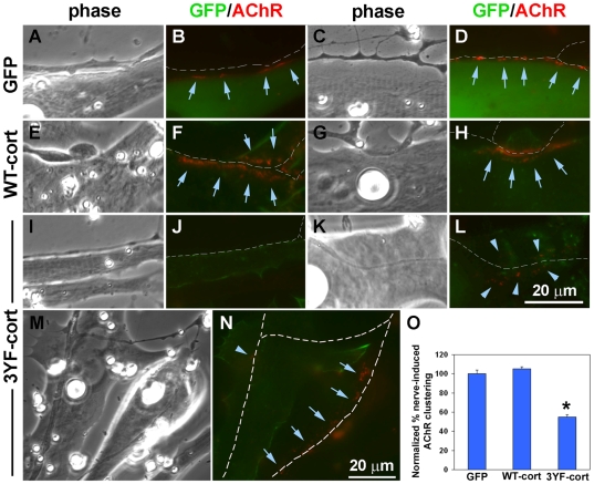
Figure 7. Suppression of synaptic AChR aggregation by phospho-mutant cortactin expressed selectively in muscle cells.Xenopus embryonic muscle cells expressing GFP (A-D) and GFP-tagged wild-type cortactin (WT-cort; E-H) and phospho-mutant cortactin (3YF-cort; I-N) were co-cultured with spinals neurons for 1 d and then labeled with R-BTX to visualize AChR clusters. Cells expressing the exogenous proteins fluoresced green and AChR clusters appeared red, as shown in this figure with 2-3 representative examples of nerves contacting muscle cells with GFP or GFP-tagged cortactin proteins. In the GFP and WT-cort muscle cells (A-H), AChRs were tightly clustered (arrows) along nerve-contacts identified (traced in white in colored panels) but this was not the case in 3YF-cort cells where nerves often induced no AChR clustering (J) or induced few clusters that were loosely organized (L; arrowheads). We found cases where the same neurites moved across normal muscle cells and 3YF-cells (M-N) and in such cases synaptic AChR clustering was robust in the normal cells (arrows) but not mutant cells (arrowheads). O. The percentages of nerve-contacts with AChR clusters were determined by examining several co-cultures with muscle cells expressing GFP or the GFP-tagged cortactin proteins (see Methods) and these values were normalized relative to numbers obtained from examining nerve-contacts on GFP-cells. In muscle cells expressing phospho-mutant cortactin, synaptic AChR clustering was almost halved. Nerve-muscle contacts examined: GFP cells, 168; WT-cort cells, 214; 3YF-cort cells, 225; mean and SEM shown, *P<0.0001. Image published in: Madhavan R et al. (2009) Madhavan et al. Creative Commons Attribution license Larger Image Printer Friendly View |
