XB-IMG-154405
Xenbase Image ID: 154405
|
|
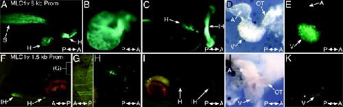
Figure 3. The activity of the Xenopus MLC1v gene promoter in founder generation transgenic tadpoles. AâE: Enhanced green fluorescent protein (EGFP) expression directed by the entire 8-kb MLC1v promoter fragment (AâE) and that resulting from a 1.5-kb proximal MLC1v promoter fragment produced by PacI digestion (FâK). A: Right-lateral view of a stage 45 tadpole showing strong EGFP fluorescence in the heart, mouth, and somites. The somitic fluorescence observed in founder generation, transient-transgenics differs from that of the f1 generation transgenic line and also the endogenous MLC1v RNA expression (see Results section). This ectopic somitic expression is likely the result of nonintegrated, linear-plasmid DNA. B,C: Higher magnification right-lateral view (B) of the heart and ventral view (C) of the same tadpole photographed in A. This animal has an inverse laterality of its visceral organs, as indicated by the direction of heart looping and left-sided gall bladder location. Such situs inversions are frequently observed in laboratory stocks of Xenopus laevis. D,E: The heart, having been dissected from a stage 43 tadpole (D, brightfield image; E, darkfield image) revealing chamber myocardium-restricted EGFP fluorescence. FâK: The 1.5-kb proximal MLC1v promoter fragment drives weak EGFP expression in the same tissues as the full-length promoter, but with a patchy, mosaic distribution, as shown by the stage 46 tadpole depicted. F: Left-lateral view. G,F: High-magnification detail image of a somite (G), whose relative position in the trunk is also indicated by the white box in F. H: Ventral view of the heart. I: Ventral view of the tadpole head and body. J,K: Dissected heart (J, brightfield image) showing patchy ventricular EGFP fluorescence (K, darkfield image). The darkfield images presented in this and all subsequent figures had their input/output levels for the different color channels adjusted individually in Adobe Photoshop. This approach enables the silhouette of the animal to be observed, while giving the EGFP fluorescence a false, blueâgreen color. IH, interhyoid facial muscle; H, heart; S, somites; GB, gall bladder; A, atria; V, ventricle; OT, outflow tract; MLC, myosin light chain. Image published in: Smith SJ et al. (2005) Copyright © 2005. Image reproduced with permission of the Publisher, John Wiley & Sons. Larger Image Printer Friendly View |