XB-IMG-117881
Xenbase Image ID: 117881
|
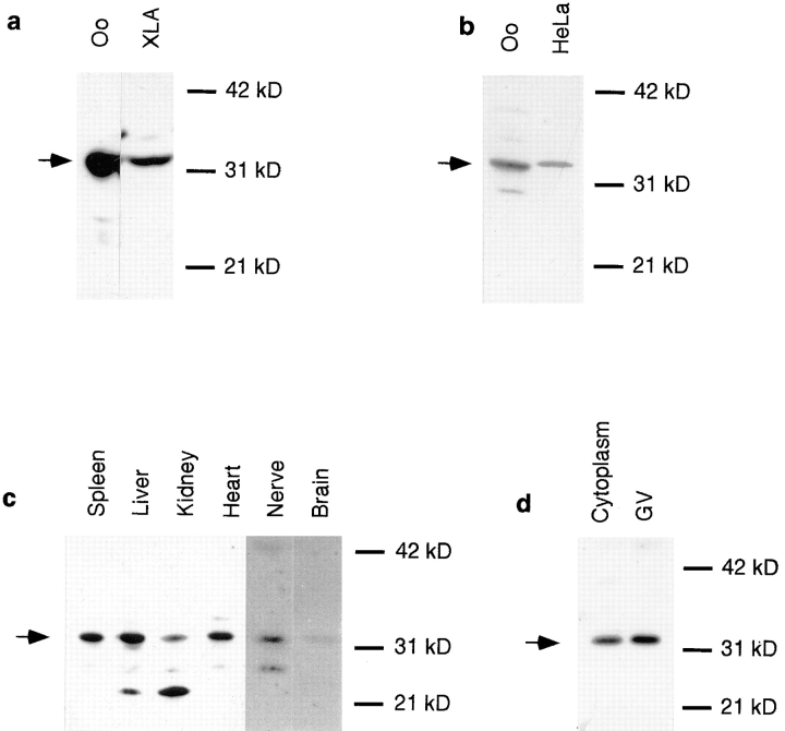
Figure 2. Distribution of Xlrbpa and TRBP in various frog tissues and HeLa cells. (a) Equal amounts of protein extract from
Xenopus oocytes (Oo) and XlA6 tissue culture cells (XLA) were
run on a SDS gel, blotted onto Immobilon membrane and detected with anti-Xlrbpa antiserum Rb6. In both tissues, a single
band of 33 kD is detectable. (b) Detection of putative human
TRBP by polyclonal serum Rb6. Oocyte extracts (Oo) and HeLa
tissue culture cell extracts (HeLa) were probed by Western blotting with polyclonal serum Rb6. Antiserum Rb6 detects Xlrbpa
in Xenopus oocytes and a band of slightly larger molecular weight
in HeLa cells representing the putative homologue, human TRBP.
(c) Distribution of Xlrbpa in various frog tissues. Equal amounts
of protein from spleen, liver, kidney, heart, nerve, and brain were
probed for the presence of Xlrbpa by Western blotting with serum Rb6. Xlrbpa can be detected in all tissues but in different
concentrations. Spleen and liver have the highest, heart and kidney intermediate, and nerve and brain lowest concentration of
Xlrbpa. In some tissues (liver, kidney) prominent degradation
products are visible. The exposure time for nerve and brain lanes
was twice that of the other tissues. (d) Intracellular distribution of
Xlrbpa. A single enucleated oocyte (Cytoplasm) and 20 isolated
nuclei (GV) were separated on an SDS gel and probed by Western blotting with serum Rb6. About equal amounts of Xlrbpa can
be detected in both lanes. Since a GV occupies <10% of the total
oocyte volume, the proteins in the cytoplasmic lanes are derived
from half the volume than in the GV lanes. The concentration of
Xlrbpa in the nucleus is, therefore, about half of that in the cytoplasm. Arrows indicate position of Xlrbpa in all gels. Image published in: Eckmann CR and Jantsch MF (1997) Image reproduced on Xenbase with permission of the publisher and the copyright holder. Creative Commons Attribution-NonCommercial-ShareAlike license Larger Image Printer Friendly View |