XB-IMG-126507
Xenbase Image ID: 126507
|
|
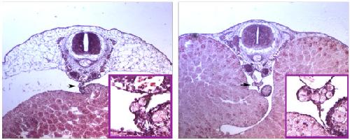
Figure 2. PGCs in embryo sections. (A) PGCs are present in the dorsal mesentery (arrow) which attaches the yolky endoderm to the rest of the body of this TS8 embryo. The inset shows an enlargement with three PGCs in the mesentery. (B) At TS9, yolk-filled PGCs are clumped in the prospective genital ridges (arrow), ventral to the dorsal aorta. The inset shows an enlargement of this region. Image published in: Elinson RP et al. (2011) Copyright ©2011 Elinson et al; licensee BioMed Central Ltd. Creative Commons Attribution license Larger Image Printer Friendly View |