XB-IMG-124621
Xenbase Image ID: 124621
|
|
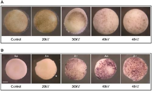
Figure 6. Whole-mount TUNEL assay exposes apoptotic cells in irradiated embryos.A. Pre-MBT embryos were exposed to different energies 20 kV, 30 kV, 40 kV and 48 kV to equal a total dose of ∼65 Gy. Non-irradiated embryos are referred to as “control”. Six hours after the MBT, embryos were fixed in MEMFA as described in the “Materials and Methods” section and photographed. B. TUNEL staining was performed on fixed embryos to detect DNA fragmentation. Embryos were treated as described in (A). Intense TUNEL staining was detected in the animal pole portion of the embryos. The embryos shown in B are representative of the TUNEL staining observed following analysis of ∼80 embryos of which 20% were stained. Arrowhead points to labeled nuclei. An, animal pole. Scale bar, 250 µm. Image published in: Dong J et al. (2010) Dong et al. Creative Commons Attribution license Larger Image Printer Friendly View |