XB-IMG-127920
Xenbase Image ID: 127920
|
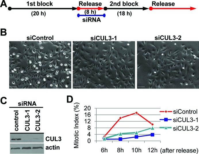
Fig. 2. CUL3 functions in mitotic entry.(A) A schematic protocol for cell synchronization (by double thymidine block) and RNAi. (B, C, D) U2OS cells were synchronized and transfected as indicated and subjected to the protocol shown in (A) with indicated siRNAs. (B) Phase pictures of indicated siRNA transfected cells at 8 hours after release. Scale bars, 100 μm. (C) When released from 2nd thymidine block, cells were lysed and subjected to WB with indicated antibodies. (D) Cells were fixed and stained with DAPI at the indicated times after release. Over 500 cells were analyzed and mitotic index was determined. Image published in: Moghe S et al. (2012) © 2011. Creative Commons Attribution-NonCommercial-ShareAlike license Larger Image Printer Friendly View |