XB-IMG-175334
Xenbase Image ID: 175334
|
|
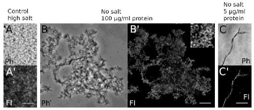
Figure 6. Light microscopy reveals reticular HisMyc-NE81ΔNLSΔCLIM assemblies. Purified protein was fixed with formaldehyde and stained with anti-NE81/anti-rabbit-AlexaFluor 488. Phase-contrast (A–C; Ph) and fluorescence images (A–C′; Fl) are shown. (A,A′) At high NaCl concentration, no clear assemblies are visible, whereas the protein forms reticular assemblies at no-salt conditions and high protein concentration (B,B′). At low protein concentration (C,C′), individual filamentous structures become apparent. Scale bars = 5 µm (inset = 1 µm). Image published in: Grafe M et al. (2019) © 2019 by the authors. Creative Commons Attribution license Larger Image Printer Friendly View |