XB-IMG-126581
Xenbase Image ID: 126581
|
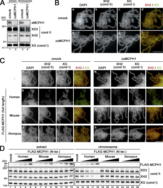
Figure 4. Characterization of xMCPH1 and mMCPH1 in the cell-free assay. (A) Sperm chromatin was added to metaphase egg extracts that had been depleted with control IgG (Δmock; lanes 1 and 3) or anti-xMCPH1 (ΔxMCPH1; lanes 2 and 4). After incubation for 120 min, chromosomes were isolated, and their associated polypeptides (lanes 3 and 4) and aliquots of the extracts (lanes 1 and 2) were analyzed by immunoblotting using the antibodies indicated. (B) Metaphase chromosomes were assembled in the mock-depleted (Δmock; a–d) or xMCPH1-depleted (ΔxMCPH1; e–h) extracts, fixed, and double stained with anti–XCAP-H2 (XH2) and anti–XCAP-G (XG). Bulk chromosomal DNA was counterstained with DAPI. (C) A reticulocyte lysate containing no hMCPH1 (−) or FLAG-tagged full-length MCPH1 from human, mouse, and Xenopus was mixed with 10 vol metaphase egg extracts that had been depleted with control IgG (Δmock; rows a–d) or anti-xMCPH1 (ΔxMCPH1; rows e–h) and incubated for 30 min. Sperm chromatin was then added and incubated for another 120 min. The resulting metaphase chromosomes were analyzed as in B. (D) Increasing concentrations of the N-terminal (N-ter.) domains of human, mouse, and Xenopus MCPH1 were mixed with metaphase egg extracts (lanes 1–13) and tested for their ability to inhibit chromosomal loading of condensin (cond) II. The relative concentrations of MCPH1 added were 1:8 (lanes 2, 6, and 10), 1:4 (lanes 3, 7, and 11), 1:2 (lanes 4, 8, and 12), and 1 (lanes 5, 9, and 13); the highest concentration roughly corresponded to the standard one used in other experiments. Sperm chromatin was incubated with these extracts, and chromosome-bound fractions (lanes 14–26) were isolated and analyzed by immunoblotting. Bars, 5 µm. Image published in: Yamashita D et al. (2011) © 2011 Yamashita et al. Creative Commons Attribution-NonCommercial-ShareAlike license Larger Image Printer Friendly View |