XB-IMG-116740
Xenbase Image ID: 116740
|
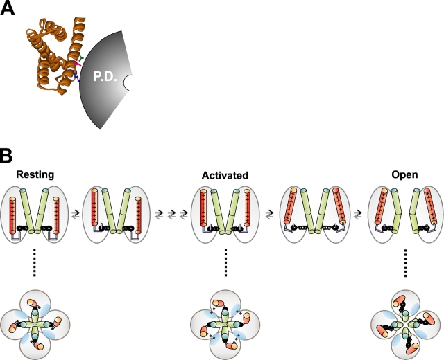
Figure 8. . A model for channel opening. (A) Mapping of Shaker ILT positions onto the crystal structure of KvAP's isolated voltage sensing (S1–S4) domain (Jiang et al., 2003a) shows ILT residues to face away from other membrane segments in direction deduced to interact with the pore domain in the activated conformation (Gandhi et al., 2003). ILT residues are colored. P.D., pore domain. (B) Model for association between activation and gating motions of S4 and opening of the S6 gate. Top layer shows a side view of two subunits of the channel; dotted lines point to a top view of the channel with all four subunits shown. The S4s (orange cylinders) move independently (or with a mild cooperativity) of each other during activation, and do not exert force on the S6 gate (green cylinders). In the activated state, each S4 interacts with its neighboring S5 (blue oval, top view), which is denoted by dashed line. From the activated state, the S4s undergo a cooperative motion in which each S4 places strain on the S6 gate via S4–S5 (hook and spring representation). The interaction of an S4 with its neighboring S5 is stronger in ILT, hence a strong depolarization is needed to release S4 from its activated conformation to undergo its final motion. Image published in: Pathak M et al. (2005) Copyright © 2005, The Rockefeller University Press. Creative Commons Attribution-NonCommercial-ShareAlike license Larger Image Printer Friendly View |