XB-IMG-121127
Xenbase Image ID: 121127
|
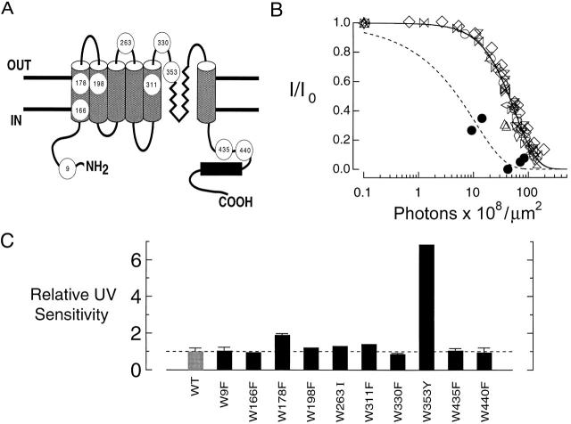
Figure 12. UV sensitivity of CNG channel tryptophan mutants. (A) Schematic diagram of RET channel α subunit indicating the positions and presumed locations of the 10 tryptophan residues in each subunit of the bovine retinal cGMP-gated channel. (B) UV dose–response relations of RET channel tryptophan point mutants. Each type of symbol represents collected results for one mutant. Mutations, D1/2 values in photons · 108 · μm−2, and the corresponding symbols are: W9F, 43.9 (○); W166F, 47.7 (□); W178F, 24.1 (▵); W198F, 37.4 (▿); W263I, 35.1 (wide diamond); W311F, 32.6 (tall diamond); W330F, 52.8 (⋄); W353Y, 6.7 (•); W435F, 43.3 (▹◃); and W440Y, 47.8 (hourglass). The dashed curve is a fit to the results for W353Y channels using ; the solid curve is a fit of this model to the collected results for all other mutants. Channels were irradiated in the absence of ligand at 280 nm. (C) Comparison of UV sensitivities of wild-type and tryptophan mutant channels. UV sensitivities, relative to the value for wild-type (WT) RET channels, are shown on the ordinate for each tryptophan point mutant. Image published in: Middendorf TR et al. (2000) © 2000 The Rockefeller University Press. Creative Commons Attribution-NonCommercial-ShareAlike license Larger Image Printer Friendly View |