XB-IMG-159744
Xenbase Image ID: 159744

|
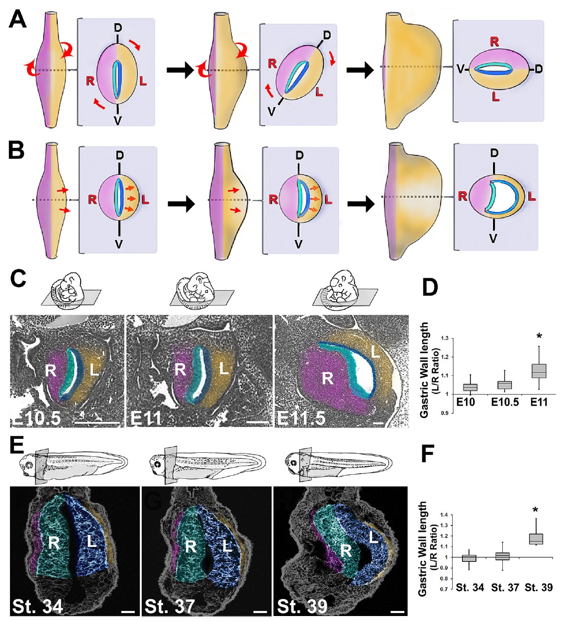
Fig. 1. Early stomach undergoes leftward expansion. The rotation model (A) posits that the embryonic stomach (shown in ventral views and cross-sections at successive stages) rotates around its longitudinal axis, shifting its dorsal face leftward. An alternative model (B) theorizes that the left wall expands more than the right. Sections of E10.5, E11 and E11.5 mouse embryos (C) or stage 34, 37 and 39 frog embryos (E) reveal the leftward expansion of the early stomach. The left/ right ratio of the lengths of the stomach walls becomes significantly greater than 1 in mouse by E11 (D) and in frog by stage 39 (F); *P<0.05. Sections in C and E are false-colored to match diagrams in A and B, highlighting layers of the stomach: right mesoderm, pink; right endoderm, teal; left endoderm, blue; left mesoderm, gold. In all sections, dorsal is upwards and the left side of animal is on right side of image. D, dorsal; V, ventral; L, left; R, right. Scale bars: 500 μm in C (E11.5, 150 μm); 75 μM in E. Image published in: Davis A et al. (2017) Copyright © 2017. Image reproduced with permission of the Publisher and the copyright holder. This is an Open Access article distributed under the terms of the Creative Commons Attribution License. Larger Image Printer Friendly View |
