XB-IMG-128499
Xenbase Image ID: 128499
|
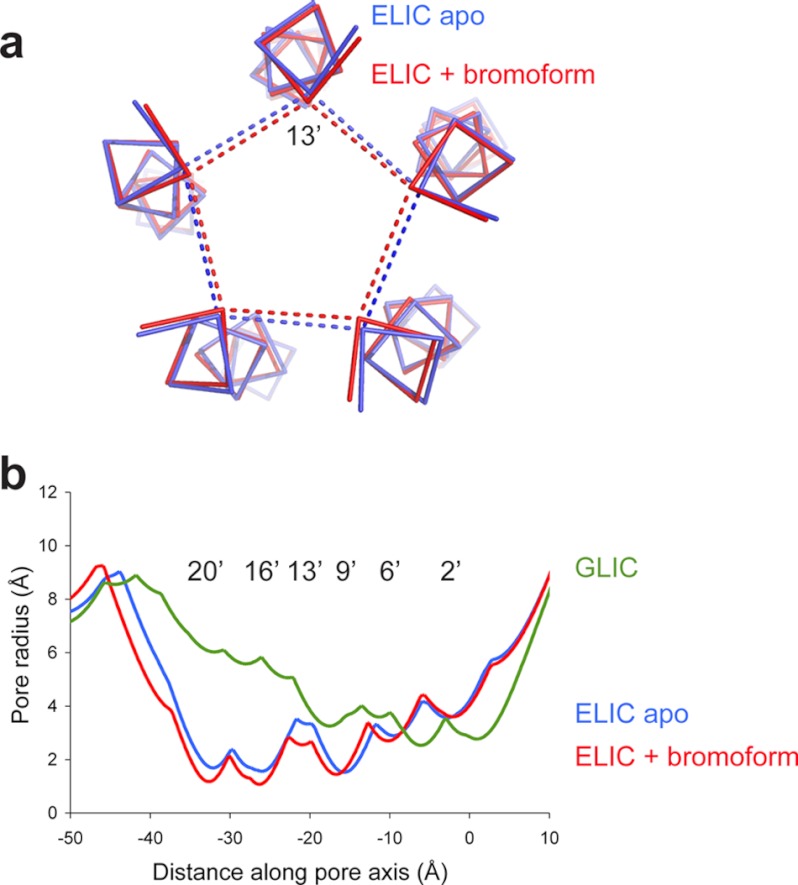
FIGURE 3. Bromoform stabilizes the ELIC pore in a closed conformation.
a, ribbon representation of the pore-lining M2-helix in the apoELIC structure (blue, PDB code 2vl0) and the bromoform-bound structure (red). The view is along the five-fold symmetry axis looking down on the channel pore from the extracellular domain. The dashed lines are distance measurements between 13′A Cα atoms of different subunits. b, pore radius analysis for apoELIC (blue), bromoform-bound ELIC (red), and GLIC, which likely corresponds to an open pore conformation (PDB code 3eam). Image published in: Spurny R et al. (2013) © 2013 by The American Society for Biochemistry and Molecular Biology, Inc. Creative Commons Attribution-NonCommercial license Larger Image Printer Friendly View |