XB-IMG-124995
Xenbase Image ID: 124995
|
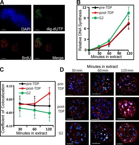
Figure 2. Asynchronous rereplication of chromocenters in G2 phase. (A) Geminin prevents in vitro replication of G2 phase chromatin. G2 phase nuclei from mouse C127 cells synchronized and prelabeled with BrdU as described in Fig. 1 were introduced into a Xenopus egg extract supplemented with geminin and digoxigenin-dUTP for 120 min. Nuclei were stained with fluorescent anti-BrdU (red) and anti-dig (green) antibodies, and DNA was counterstained with DAPI (blue). BrdU-positive nuclei (still in S phase at the time of collection) incorporated digoxigenin-dUTP as a result of elongation of preexisting forks, but no initiation of replication was observed within G2 phase nuclei. (B) Replication proceeds efficiently within G1 and G2 phase nuclei. Nuclei prepared as described in Fig. 1 A were introduced into a Xenopus egg extract supplemented with digoxigenin-dUTP. Aliquots were removed at the indicated times, and nuclei were stained with fluorescent anti-dig (and for G2 phase nuclei anti-BrdU) antibodies. The incorporation of digoxigenin-dUTP per (BrdU negative) nucleus was quantified using an imaging system with softWoRx and normalized to the lowest value for all nuclei in all preparations. n > 30 nuclei per condition/time point (arbitrary units). Note that G2 phase nuclei incorporate 40% more digoxigenin-UTP. Because there is twice as much DNA in G2 phase nuclei, the efficiency of replication is ∼70% that of G1 phase. (C) Chromocenters are preferentially replicated late only in post-TDP nuclei. Nuclei prepared as described in Fig. 1 A were introduced into a Xenopus egg extract, and aliquots were removed and pulse labeled for 5 min with biotin-dUTP at the indicated times. Nuclei were stained with fluorescent avidin to highlight incorporated biotin, and chromocenters were identified as sites of intense DAPI staining. G2 phase nuclei were additionally stained with anti-BrdU antibodies as in A. Colocalization between chromocenters and in vitro biotin labeling was quantified using softWoRx. Pre-TDP and G2 phase nuclei showed a similar amount of colocalization throughout the in vitro reaction, whereas post-TDP nuclei showed significantly more chromocenter replication late in the reaction (120 min). n > 50 nuclei/condition/time point. (D) Exemplary images of individual pre-TDP, post-TDP, and G2 phase nuclei from C fixed and stained at different time points during the in vitro reaction. Colocalization between chromocenters (DAPI-dense regions in blue) and in vitro biotin-dUTP pulse labeling (red) are highlighted in white. For G2 phase, only BrdU-negative nuclei were analyzed. Note that the post-TDP 120-min nucleus shows considerably more pixels colocalized with chromocenters than other nuclei. Error bars indicate mean ± SD. Bars, 5 µm. Image published in: Lu J et al. (2010) © 2010 Lu et al. Creative Commons Attribution-NonCommercial-ShareAlike license Larger Image Printer Friendly View |