XB-IMG-129670
Xenbase Image ID: 129670
|
|
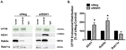
Figure 8. siSGK1 increased the amount of wt-CFTR in early endosomes, but not in recycling endosomes.Co-immunoprecipitation studies were conducted to determine the subcellular location of wt-CFTR in siNeg and siSGK1 transfected cells treated with dexamethasone (50 nM for 4 hours). EEA and Rab5a are markers of early endosomes, and Rab11a is a marker of recycling endosomes. wt-CFTR was immunoprecipitated, and co-immunoprecipitated proteins were eluted into SDS sample buffer, and separated by 7.5% SDS-PAGE. The blots were then probed for wt-CFTR, EEA, Rab5a, and Rab11. Co-immunoprecipitation of wt-CFTR with EEA1 and Rab5a identifies the amount of wt-CFTR in early endosomes, and co-immunoprecipitation of wt-CFTR with Rab11a identifies the amount of wt-CFTR in recycling endosomes. Quantification of data for Rab and EEA1 immunoprecipitation with wt-CFTR in siNeg and siSGK1 cells is normalized for the total amount of wt-CFTR immunoprecipitated. Blots in siNeg and siSGK1 experiments were cut for presentation, but were run one the same blot to allow for comparison. Lysate, the amount of EEA1, Rab5a Rab11 and wt-CFTR in cell lysates. CFTR IP, indicates IP with the anti-CFTR antibody and then western blot with the indicated antibody. IgG, immunoprecipitation with a non-specific antibody. (A) Representative western blots and (B) Summary of the data. Experiments repeated three times. *P<0.05 versus siNeg. Image published in: Bomberger JM et al. (2014) Image reproduced on Xenbase with permission of the publisher and the copyright holder. Creative Commons Attribution license Larger Image Printer Friendly View |