XB-IMG-126140
Xenbase Image ID: 126140

|
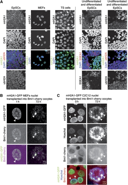
Figure 7. mH2A correlates with stable Xi and is maintained after nuclear transfer to Xenopus oocytes. (A) Immunostaining of female EpiSCs, MEFs and TS cells against mH2A1 (green), ubH2A (red) and SSEA1 (red). Undifferentiated female EpiSCs do not exhibit accumulation of mH2A1 on the ubH2A-labelled Xi. mH2A1 is induced in differentiated EpiSCs, marked by loss of the pluripotency marker SSEA1 (right panel, left column). mH2A1 is incorporated in chromatin of the Xi in differentiated EpiSCs, as shown by co-localization with Xi marker ubH2A (right panel, right column). Note that the Xi of undifferentiated EpiSCs is stained with ubH2A only. mH2A1 was found accumulated in 95% of female MEFs, 85% of female TS cells and 0% of female EpiSCs. DAPI is shown in blue. Scale bars=10 μm. Images are projected Z-sections. (B, C) mH2A1-GFP remains associated with heterochromatic regions in transplanted nuclei and reveals chromatin reorganization. Projections of confocal images of mH2A1-GFP MEF (B) and sable mH2A1-GFP C2C12 (C) nuclei transplanted into oocytes preloaded with Bmi1-cherry by mRNA injection. Note the persistence of mH2A1-GFP on the Xi (arrowheads), bound by Bmi1-cherry imported from the oocyte, and the appearance of mH2A1-GFP-labelled pericentric heterochromatin foci. Scale bars=10 μm. Images are projected Z-sections. Image published in: Pasque V et al. (2011) Copyright © 2011, European Molecular Biology Organization. Creative Commons Attribution-NonCommercial-ShareAlike license Larger Image Printer Friendly View |
