XB-IMG-117365
Xenbase Image ID: 117365
|
|
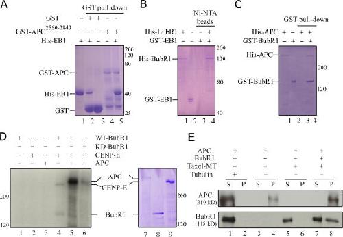
Figure 6. BubR1 phosphorylates APC and forms a ternary complex with APC and microtubules in vitro. (A–C) In vitro binding assay using purified proteins as indicated. Purified recombinant proteins were incubated with glutathione–Sepharose 4B (A and C) or Ni-NTA agarose (B) beads as indicated. Protein pulldown with the beads was assayed by SDS-PAGE and Coomassie blue staining. (D) APC phosphorylation by BubR1. In vitro kinase assay was performed with a combination of purified recombinant APC (lane 7), BubR1 (lane 8), and CENP-E (lane 9) as indicated. (E) BubR1, APC, and microtubules form a ternary complex. After centrifugation through a 40% sucrose cushion, BubR1–APC–taxol microtubule complex formation was assayed by immunoblotting. S, supernatant; P, pellet. Image published in: Zhang J et al. (2007) Copyright © 2007, The Rockefeller University Press. Creative Commons Attribution-NonCommercial-ShareAlike license Larger Image Printer Friendly View |