XB-IMG-127272
Xenbase Image ID: 127272
|
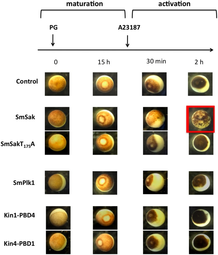
Figure 5. SmSak induces de novo centriole formation in in vitro matured Xenopus oocytes.Samples of cRNA encoding SmSak, SmSakT175A, SmPlk1 or Kin1-PBD4 and Kin4-PBD1 hybrid proteins, were microinjected in oocytes and incubated for 2 h for protein expression, before addition of PG. At 15 h following the addition of PG, GVBD monitored by the formation of a white spot centered at the germinal pole of Metaphase II arrested oocytes, was observed in all oocyte groups. Matured oocytes were then activated with calcium ionophore A23187, and surface morphology was observed after 30 min or 2 h. Only SmSak-expressing oocytes formed pigmented speckles that increased in number from 30 min to 2 h. In all other oocytes, the animal pole gradually darkened after the addition of A23187 and the formation of a black spot (fertilization coat) could be seen. Image published in: Long T et al. (2012) Long et al. Creative Commons Attribution license Larger Image Printer Friendly View |