XB-IMG-127344
Xenbase Image ID: 127344
|
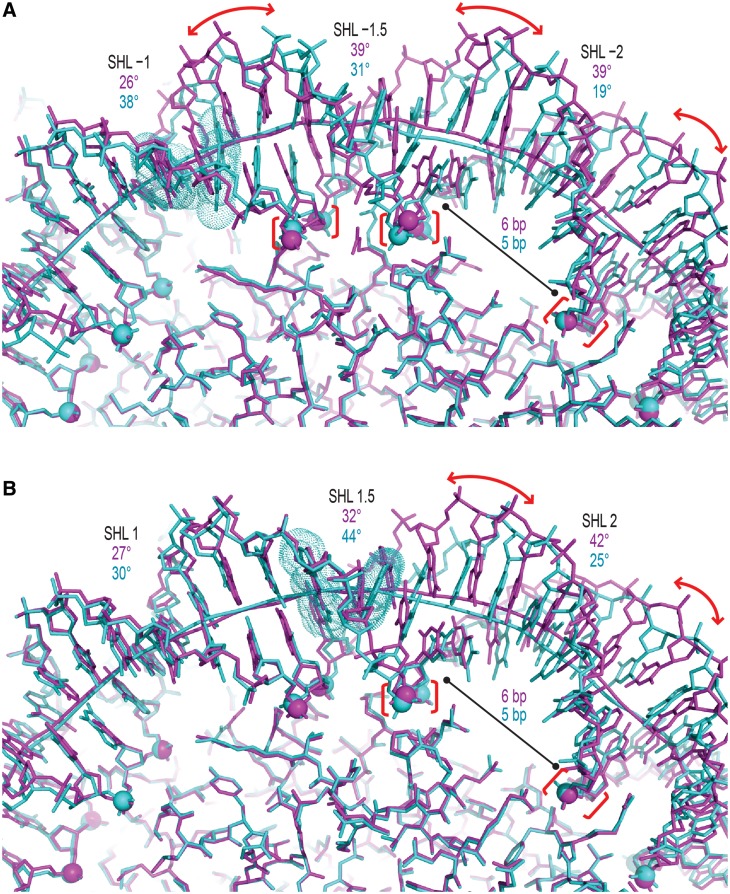
Figure 6. DNA single-strand histone attachment points act as hinges to allow double helix conformational freedom. (A and B) DNA stretching around SHL ± 1 to SHL ± 2 in NCP145 (cyan) gives rise to substantial structural differences compared to NCP147 (magenta), which displays no stretching but is composed of a DNA with nearly identical sequence. Histone association with the binding platforms (phosphorous atoms, spheres) is very similar between the two constructs, whereby the extreme stretch-associated kinking in NCP145 (bases, space-filling dots) at either SHL −1 (A; CA = TG, roll = 38°, rise = 5.0 Å) or SHL 1.5 (B; GG = CC, roll = −52°, rise = 5.7 Å) is accommodated largely by swivel-like repositioning of the double helix (curved arrows) about the hinges (brackets). This results in a distinct distribution of helix axis (tubes) curvature between the major and minor groove-inward regions (values shown). Image published in: Chua EY et al. (2012) © The Author(s) 2012. Creative Commons Attribution-NonCommercial license Larger Image Printer Friendly View |