XB-IMG-125933
Xenbase Image ID: 125933
|
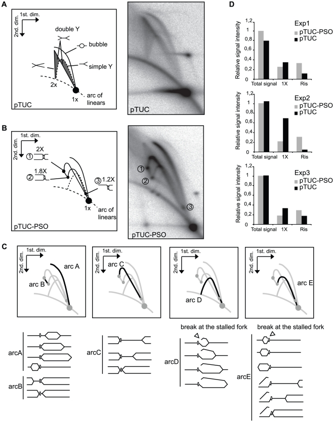
Figure 2. Two-dimensional gel electrophoretic analysis of pTUC and pTUC-PSO plasmids replicating in Xenopus egg extracts.Plasmids pTUC (A) and pTUC-PSO (B) were incubated 95 min in Xenopus egg extract in the continuous presence of [α-32P]-dATP. Plasmids were purified, linearized by AflIII digestion and analyzed by 2D gel electrophoresis. Phosphorimager images of the dried gels and interpretative diagrams are shown. (C) Interpretation of the pTUC-PSO specific arcs. (D) Relative intensity of total signal, 1x spot and replication intermediates (RIs) from 3 independent experiments (Exp1, Exp2, Exp3). Image published in: Le Breton C et al. (2011) Le Breton et al. Creative Commons Attribution license Larger Image Printer Friendly View |