XB-IMG-122577
Xenbase Image ID: 122577
|
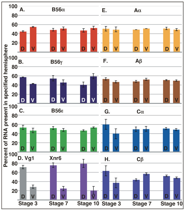
Figure 7. The dorsoventral distribution of PP2A subunits in Xenopus embryos. Xenopus laevis embryos at stages 3, 7, and 10 were hemisected into dorsal and ventral halves. RNA was purified from the hemisected halves and real time RT-PCR was carried out. The data were normalized to ODC expression levels, and are presented as the percentage of the total signal present in the specified hemisphere. PP2A A and C subunits Aα (E), Aβ (F), Cα (G), and Cβ (H) are relatively equally distributed dorsoventrally or slightly enriched dorsally. However, B56α (A) at stage 3 and B56γ (B) at stage 10 are more abundant ventrally. Dissection controls Vg1 (stage 3) and Xnr6 (stages 7 and 10) are enriched dorsally (D). D = dorsal, V = ventral. Image published in: Baek S and Seeling JM (2007) Copyright © 2007 Baek and Seeling; licensee BioMed Central Ltd. Creative Commons Attribution license Larger Image Printer Friendly View |