XB-IMG-171095
Xenbase Image ID: 171095

|
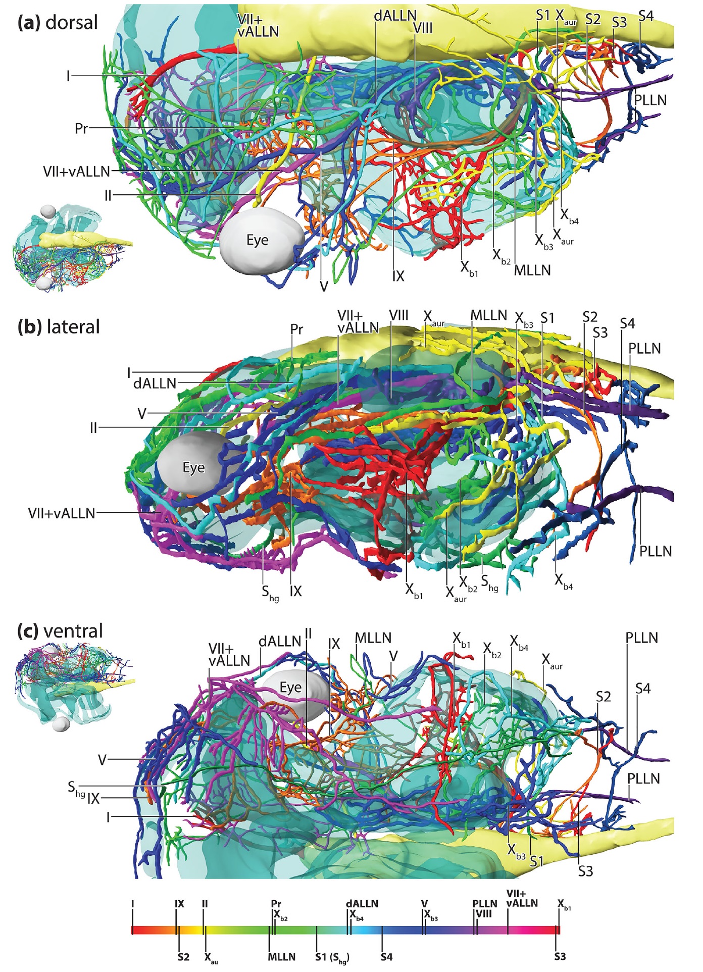
FIGURE 2 3D reconstruction of a dorso-ventral scan of a whole mount antibody staining against acetylated-alpha tubulin of a Xenopus laevis
larva at NF stage 47/48. The nerves have been reconstructed only on the left side. Cranial nerves are color-coded according to a rainbow
color map. The brain is shown in light yellow, the eye in white, and the cartilaginous head skeleton in transparent blue. A smaller
overview of the whole 3D model is shown in the lower left corner. (b) lateral view. (c) ventral view. A smaller overview of the whole 3D
model is shown in the upper left corner [Color figure can be viewed at wileyonlinelibrary.com] Image published in: Naumann B and Olsson L (2018) Copyright © 2018. Image reproduced with permission of the Publisher, John Wiley & Sons. Larger Image Printer Friendly View |
