XB-IMG-117883
Xenbase Image ID: 117883
|
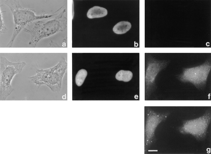
Figure 4. Localization of Xlrbpa and ribosomes in HeLa cells. Coverslip-grown HeLa cells were stained with preimmune serum Rb6 (a–
c), immune serum Rb6 (d–f), and anti-ribosomal serum 13751 (g). (a and d) Phase contrast images, (b and e) DAPI images, (c and f)
FITC images, and (g) rhodamine image. Preimmune serum Rb6 shows no signal in HeLa cells (c), while staining with immuneserum
Rb6 reveals a homogenous, slightly punctate pattern in the entire cell (f). (g) Cells in f were double-labeled with an anti-ribosomal serum 13751 which was visualized in the rhodamine channel. Staining with both anti-Xlrbpa and anti-ribosomal serum 13751 resulted in
very similar images, revealing a homogeneous, slightly punctate staining pattern over the entire cell. Bar, 10 μm. Image published in: Eckmann CR and Jantsch MF (1997) Image reproduced on Xenbase with permission of the publisher and the copyright holder. Creative Commons Attribution-NonCommercial-ShareAlike license Larger Image Printer Friendly View |