XB-IMG-125645
Xenbase Image ID: 125645
|
|
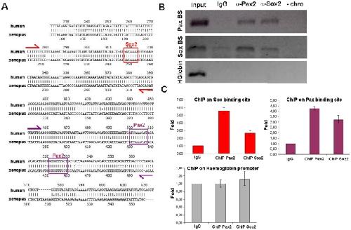
Figure 6. Pax2 and Sox2 are directly recruited to the HCNR 81675 DNA.(A) rVista 2.0 alignment of HCNR 81675 human and Xenopus genomic sequence. Conserved binding sites for Sox and Pax2/5/8 proteins found by TRANSFAC are represented. Primer location enclosing the genomic region of Sox and Pax2/5/8 binding sites designed for chromatin immunoprecipitation are marked with red and violet arrows respectively. (B) ChIP with anti-Pax2 and anti-Sox2 antibodies from stage 30–34 Xenopus otic vesicles was performed and the PCR amplification of the DNA fragments pulled down by Pax2 and Sox2 chromatin immunoprecipitation is shown. A region of the haemoglobin locus with no Pax2 and Sox2 binding sites shows no immunoprecipitation with these antibodies. (C) Graphs representing the relative fold enrichment of Sox2 and Pax2 binding to the HCNR 81675 but not to the haemoglobin region detected by quantitative PCR. Image published in: Robert-Moreno À et al. (2010) Robert-Moreno et al. Creative Commons Attribution license Larger Image Printer Friendly View |