XB-IMG-153726
Xenbase Image ID: 153726
|
Fig. 5.
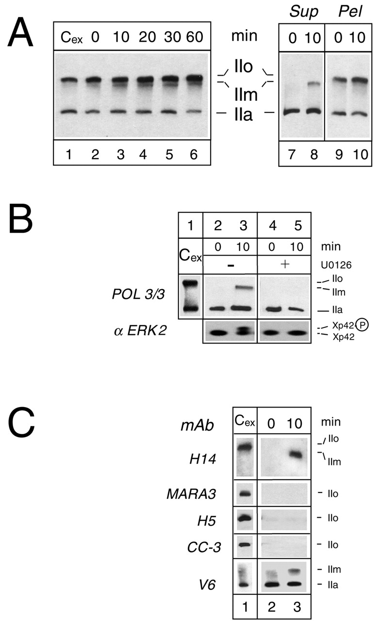
The IIm form of RPB1 is generated in serum-stimulated A6 cells. (A) Serum was added to quiescent A6 cells. Whole lysates (left) or lysates fractionated in a low-salt buffer into cytosolic supernatants (Sup) and pellets (Pel) from exponentially growing (Cex), quiescent (0) or serum-stimulated quiescent cells were analysed by western blot using the antibody POL3/3. The duration of the stimulation is indicated in minutes (min). (B) Whole lysates from exponentially growing A6 cells (Cex), cytosolic fraction from quiescent cells (0) or quiescent cells stimulated by serum during 10 minutes (10) were analysed by western blot. U0126 was added (lanes 4, 5) or not (lanes 2, 3) to culture medium 30 minutes before serum stimulation. The phosphorylation state of RPB1 and Xp42 was monitored using the POL3/3 and anti-ERK2 monoclonal antibodies (mAb), respectively. (C) Immunoreactivity of the IIm form. The samples from lanes 1 to 3 used in Fig. 5B were analysed by western blot with the indicated monoclonal antibodies. The positions of the IIa, IIo and IIm forms are indicated. Image published in: Palancade B et al. (2001) Copyright © 2001. Image reproduced with permission of the Publisher, The Company of Biologists Ltd. Larger Image Printer Friendly View |