XB-IMG-129076
Xenbase Image ID: 129076

|
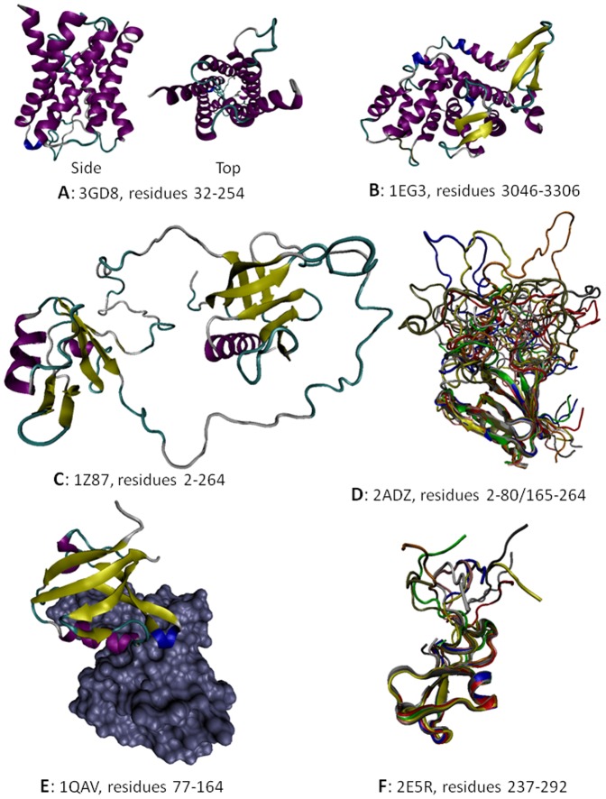
Figure 4. Structural information on some members of the dystrophin-associated protein complex: A. Crystal structure of the human AQP-4 fragment (PDB ID: 3GD8, residues 32–254); B. Crystal structure of the human dystrophin fragment (PDB ID: 1EG3, residues 3046–3306).C. NMR solution structure of the mouse α-1 syntrophin fragment (PDB ID: 1Z87, residues 2–264 that correspond to the PHN–PDZ–PHC module). D. NMR solution structure of the mouse α-1 syntrophin fragment (PDB ID: 2ADZ, residues 2–80/165–264 that correspond to the PHN-‘L’-PHC construct). Ten representative members of the conformational ensemble are shown by chains of different color. E. Crystal structure of a complex (PDB ID: 1QAV) between the PDZ domain of mouse α-1 syntrophin (residues 77–164, shown as a colored chain) and the Neuronal nitric oxide synthase, nNOS (shown as gray surface). F. NMR solution structure of the fragment of human α-dystrobrevin (PDB ID: 2E5R, residues 237–292 that correspond to the ZZ-domain). Ten representative members of the conformational ensemble are shown by chains of different color. Image published in: Na I et al. (2013) Image reproduced on Xenbase with permission of the publisher and the copyright holder. Creative Commons Attribution license Larger Image Printer Friendly View |
