XB-IMG-123008
Xenbase Image ID: 123008
|
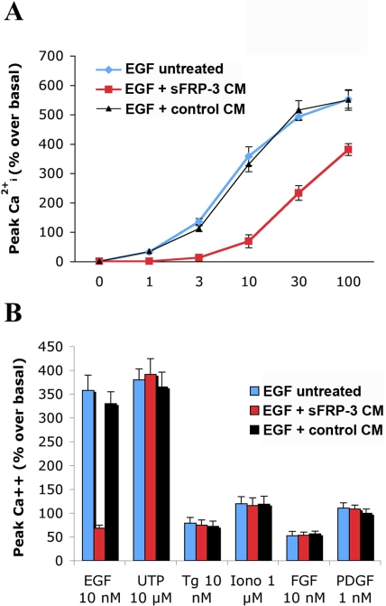
Figure 4. sFRP-3 inhibits the EGF-induced [Ca2+]i variations in EGFR-T17 cells.Fura-2 loaded cells, suspended in KRH medium, were incubated with medium conditioned (CM) either by sFRP-3-expressing or not expressing cells, and challenged with increasing concentrations of EGF (A), or submaximal concentrations of FGF (10 nM), PDGF (1 nM), UTP (10 µM), thapsigargin (Tg, 10 nM) or iomomycin (Iono, 1 µM) as indicated (B). Shown are the [Ca2+]i changes±s.e.m. measured as % increases over resting [Ca2+]i values (n = 4). These were 136±12 nM and were not changed by the addition of sFRP-3 CM or control CM. Image published in: Scardigli R et al. (2008) Scardigli et al. Creative Commons Attribution license Larger Image Printer Friendly View |