XB-IMG-123192
Xenbase Image ID: 123192
|
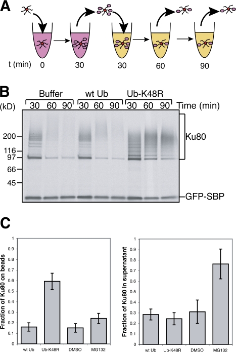
Figure 3. Polyubiquitylation induces the loss of Ku80 from DNA. (A) The experimental scheme of the release assay. At time 0, SB-DNA beads and streptavidin-coated beads were added to egg extract containing 35S-labeled Ku80 and either ubiquitin storage buffer, 0.5 mg/ml ubiquitin, 0.5 mg/ml ubiquitin-K48R, DMSO, or 100 μM MG132. Beads were incubated for 30 min, removed from the extract, and subsequently washed and incubated in extract containing the additional factors but lacking the radioactive proteins for an additional 30 or 60 min. As a control for bead recovery, we used the streptavidin beads coated with 35S-labeled GFP fused to the streptavidin-binding peptide (GFP-SBP). (B) The results of the experiment described in A after exposure to film. Extract contained buffer, 0.5 mg/ml ubiquitin, or 0.5 mg/ml ubiquitin-K48R. Polyubiquitylated Ku80 and GFP-SBP, added as a recovery control, are indicated. (C) Extract contained buffer, 0.5 mg/ml ubiquitin, 0.5 mg/ml ubiquitin-K48R, DMSO, or 100 μM MG132. Beads isolated after 30 min in extract containing radioactive Ku80 (t = 30 min), along with beads chased for an additional 30 min in nonlabeled extract (t = 60 min), were washed, and retained radioactivity was quantified using a liquid scintillation counter. In addition, samples were taken from the supernatant of the 60-min time point and precipitated onto glass microfiber filters with 10% TCA, and radioactivity was quantified using a liquid scintillation counter. The values of Ku80 remaining on beads and Ku80 in supernatant at t = 60 min were normalized relative to the radioactivity on beads at t = 30 min for each sample. Error bars denote one standard error of the mean; n = 3. Image published in: Postow L et al. (2008) © 2008 Postow et al. Creative Commons Attribution-NonCommercial-ShareAlike license Larger Image Printer Friendly View |