XB-IMG-124177
Xenbase Image ID: 124177
|
|
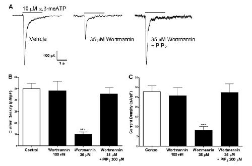
Figure 1. Sensitivity of native P2X3 receptor activity to PIP2 depletion in DRG neurons. A) Typical traces of P2X3 response to 10 μM α,β-meATP in DRG nociceptors (left), to 10 μM α,β-meATP after 2 h incubation with wortmannin (middle), to 10 μM α,β-meATP after 2 h incubation with wortmannin and intracellular application of 200 μM PIP2 (right). B) Quantitative results. P2X3 responses to 10 μM α,β-meATP under control conditions, after 2 h incubation with 100 nM wortmannin, 35 μM wortmannin, or 35 μM wortmannin with 200 μM PIP2 in the pipette solution (N = 6–13). C) Quantitative results. P2X3 responses to 10 μM ATP under control conditions, after 2 h incubation with 100 nM wortmannin, 35 μM wortmannin, or 35 μM wortmannin with 200 μM PIP2 with in the pipette solution (N = 7–18). (***, P < 0.001). Image published in: Mo G et al. (2009) Copyright © 2009 Mo et al; licensee BioMed Central Ltd. Creative Commons Attribution license Larger Image Printer Friendly View |