XB-IMG-117874
Xenbase Image ID: 117874

|
|
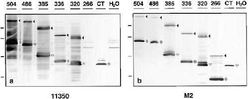
Figure 2. Western blot analysis of Xenopus oocytes injected with mRNAs corresponding to the occludin proteins diagrammed in Fig. 1. mRNAs were microinjected in the vegetal hemisphere of stage VI oocytes, and the injected oocytes were incubated at 17°C overnight. Oocyte homogenates were separated on 12% SDS-PAGE, transferred to Immobilon membranes, and then immunoblotted with either anti-occludin antibody 11350 (a) or with anti-FLAG monoclonal antibody M2 (b). The number on the top of each lane identifies each protein as named in Fig. 1. The asterisks in a and b indicate the predicted position of each expressed protein. With the exception of the soluble CT, the expressed proteins formed dimers as indicated by the arrowheads. Note that protein 266, with almost the entire COOH terminus deleted, was not recognized by 11350 antibody. H2O, control oocytes injected with water. Molecular weight markers (from top to bottom): 97.4, 66, 45, and 31 kD. Image published in: Chen Y et al. (1997) Image reproduced on Xenbase with permission of the publisher and the copyright holder. Creative Commons Attribution-NonCommercial-ShareAlike license Larger Image Printer Friendly View |
