XB-IMG-117814
Xenbase Image ID: 117814

|
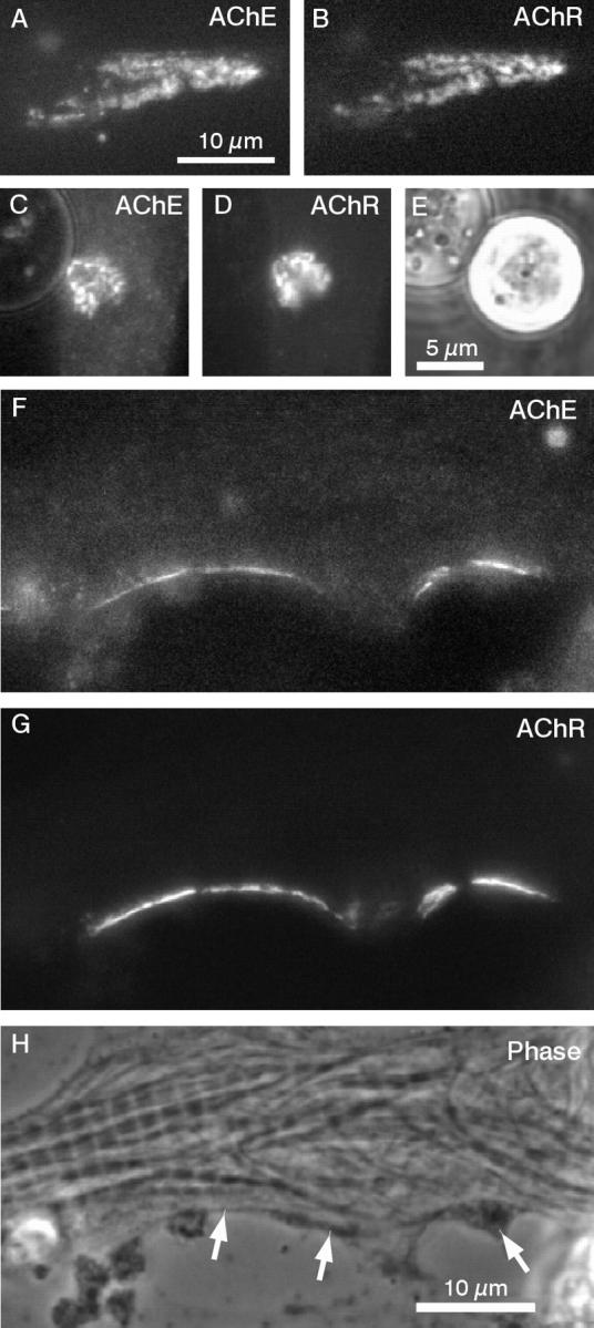
Figure 2. AChE clustering in cultured Xenopus muscle cells. (A and B) A spontaneously formed hot spot of AChE and AChR visualized by R-fasciculin 2 and OG-BTX labeling. (CâE) An AChE cluster induced by a HB-GAMâcoated bead. The culture was prelabeled with R-fasciculin 2 and OG-BTX before bead application. Thus, this cluster was formed from preexistent AChE and AChR. (FâH) Clustering of preexistent AChE at the NMJ. The muscle culture was innervated with spinal cord neurons after prelabeling with fluorescent toxins. Both AChE and AChR become clustered at sites of nerveâmuscle contact (indicated by arrows in H) formed along the length of this neurite. Image published in: Peng HB et al. (1999) Image reproduced on Xenbase with permission of the publisher and the copyright holder. Creative Commons Attribution-NonCommercial-ShareAlike license Larger Image Printer Friendly View |
