XB-IMG-159561
Xenbase Image ID: 159561
|
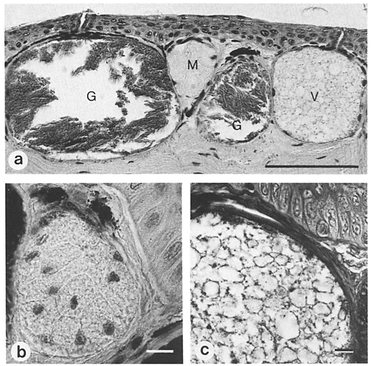
Figure 1. Light micrographs of the skin
of adult Xenopus laevis. (a) Overall view
of Bouin-fixed and azocarmin-aniline
blue stained sections show mature granular
glands (G), mucous glands (M),
and the vacuolated stage (V) of premature
granular glands. The glands lie in
the dennis and open towards the surface
via ducts. (b) Early stage in granular
gland development where the cell membranes
of the secretory cells are still intact.
(c) Detail of the vacuolated stage.
Cell membranes have now disappeared
and the nuclei are situated at the periphery
of the acinus, however, storage granules
are still absent at this stage. (a) Bar,
100 lam. (b) Bar, 10 ~tm. (c) Bar, 10 I.tm. Image published in: Flucher BE et al. (1986) Copyright © 1986. Creative Commons Attribution-NonCommercial-ShareAlike license Larger Image Printer Friendly View |