XB-IMG-128325
Xenbase Image ID: 128325
|
|
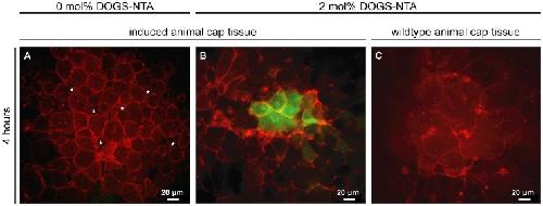
Figure 4. Animal cap tissue incubated on supported membranes: Impact of lateral density of Xcad-11.Xenopus embryos were injected in one blastomere of 2-cell stage with 300 pg tBR and 500 pg XFz7 to induce neural crest cell fate (induced animal cap tissue) or left non-induced (wildtype animal cap tissue). Additionally 500 pg GAP43-mCherry was injected as membrane tracer, and 150 pg slug-promoter-GFP as read-out for neural crest induction process. Animal cap explants were taken at stage 9 and cultivated on pure SOPC membranes (0 mol% DOGS-NTA, A) and on Xcad-11-functionalized membranes (2 mol% DOGS-NTA, B, C). Animal caps were placed on the surface and imaged after 4 hours of cultivation. Activation of slug-promoter-GFP reporter (green) indicates successful neural crest induction. NCC induced tissue started to dissociate and cells lost cell-cell contact (A, gaps are highlighted by asterisks) on pure SOPC membranes. Induced explants on Xcad-11 functionalized surface (B) consist of a cohesive tissue that shows close cell-cell contacts. A clear expression of the slug transcription factor was detected by the positive GFP signal. Wildtype animal cap tissue disintegrated and started dying (C) on functionalized membranes. Image published in: Körner A et al. (2013) Image reproduced on Xenbase with permission of the publisher and the copyright holder. Creative Commons Attribution license Larger Image Printer Friendly View |