XB-IMG-124463
Xenbase Image ID: 124463
|
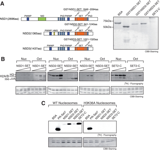
FIGURE 1. The NSD SET domain methylates H3K36 on nucleosomes. A, the left panel shows the schematic of human NSD1–3 domain organization. NID denotes the nuclear receptor interaction domain (12). PWWP denotes the PWWP domain which contains the proline-tryptophan-tryptophan-proline motif in the consensus amino acid sequence. The region of the SET domain protein expressed and used in the following enzymatic assays is indicated above the sequences. All of the proteins include the pre-SET, SET, and post-SET domain and flanking regions. The right panel shows CBB staining of the purified GST-NSD1–3 SET domain fusion proteins and the SET2 C-terminal fragment separated on SDS-PAGE. B, HKMT assay on nucleosomes (Nuc) and octamers (Oct) with enzymes analyzed in A using [3H]SAM as the methyl donor. The protein concentrations used for NSD1–3 SET were 0.05, 0.1, 0.2, and 0.4 μm; those for SET2-C were 0.01, 0.02, 0.04, and 0.08 μm. Nucleosomes and octamers contained recombinant, unmodified histones, and their final concentrations in the reactions were 0.35 μm unless otherwise noted. The assay mixtures were separated by SDS-PAGE. The lower panel shows CBB staining of the histones, and the upper panel shows results with fluorography. The asterisk denotes a proteolyzed form of H3 (see text). C, HKMT assays were performed on wild type (WT) or H3K36A nucleosomes using enzymes described in A; bovine serum albumin (BSA) and full-length recombinant PR-SET7 served as controls. The final concentrations for NSD1–3 SET in these and subsequent reactions were 0.15 μm unless otherwise noted. Image published in: Li Y et al. (2009) © 2009 by The American Society for Biochemistry and Molecular Biology, Inc. Creative Commons Attribution-NonCommercial license Larger Image Printer Friendly View |