XB-IMG-124660
Xenbase Image ID: 124660
|
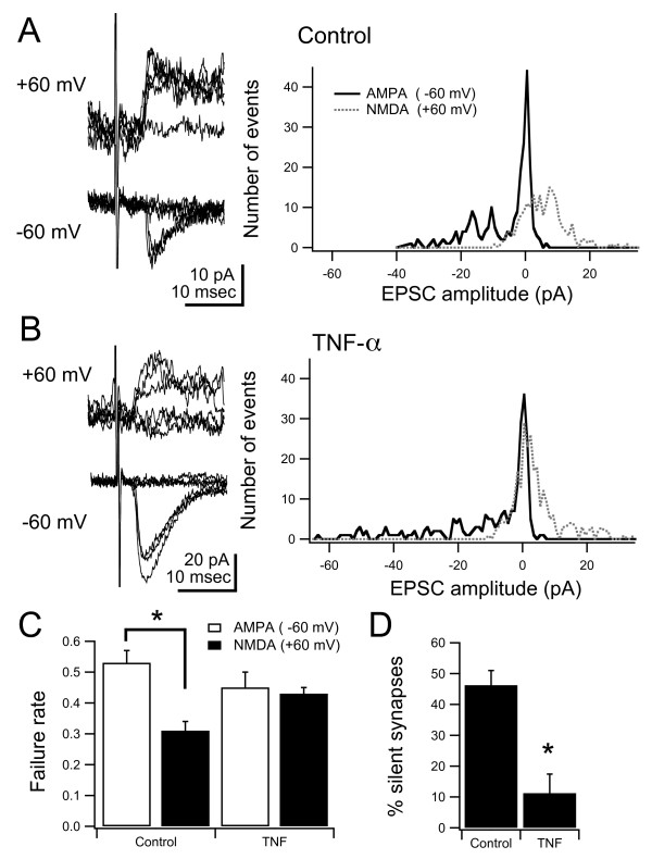
Figure 6. Long-term developmental exposure to TNF-α results in a decrease of NMDAR-only synapses. (A, B) Left: superimposed responses evoked by minimal stimulation recorded at -60 and +60 mV from control and TNF-α-treated animals. Right: histogram of all minimal stimulation AMPA and NMDA responses from control and TNF-α-treated animals. (C) Failure rates of NMDA currents were significantly lower in control animals, whereas they were not different in drug-treated animals, consistent with a decreased number of silent synapses. (D) Estimation of the percent of silent synapses in the stimulated projection was also significantly different. Asterisk indicates p < 0.05. For P-values see Results. Image published in: Lee RH et al. (2010) Copyright ©2010 Lee et al; licensee BioMed Central Ltd. Creative Commons Attribution license Larger Image Printer Friendly View |