XB-IMG-144709
Xenbase Image ID: 144709
|
|
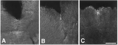
FIG. 9. Microtubule array persists in a region of new membrane delivery following cytochalasin D treatment. A–C show confocal images
of the microtubule distributions in first-cleavage embryos fixed 5, 10, and 20 min, respectively, following exposure to 10 mg/ml cytochalasin
D. In C, the surface had entirely flattened out, and the embryo resembled the live one shown in Fig. 8. Bar 50 mm. Image published in: Danilchik MV et al. (1998) Copyright © 1998. Image reproduced with permission of the Publisher, Elsevier B. V. Larger Image Printer Friendly View |