XB-IMG-118680
Xenbase Image ID: 118680

|
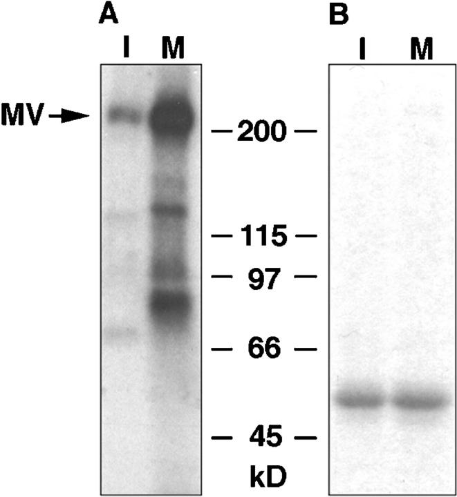
Figure 7. Phosphorylation of myosin V in cell cycle-arrested frog egg extracts. A, Extracts were 32P-labeled, and myosin V was immunoprecipitated from interphase- and metaphase-arrested extracts using affinity-purified DIL2 antibody (I and M, respectively), separated by SDS-PAGE, and analyzed by autoradiography. Myosin V (MV) is more highly phosphorylated in metaphase extracts. B, Coomassie blue stained gel as in A, to demonstrate approximately equal protein load for both treatments. Image published in: Rogers SL et al. (1999) © 1999 The Rockefeller University Press. Creative Commons Attribution-NonCommercial-ShareAlike license Larger Image Printer Friendly View |
