XB-IMG-157688
Xenbase Image ID: 157688

|
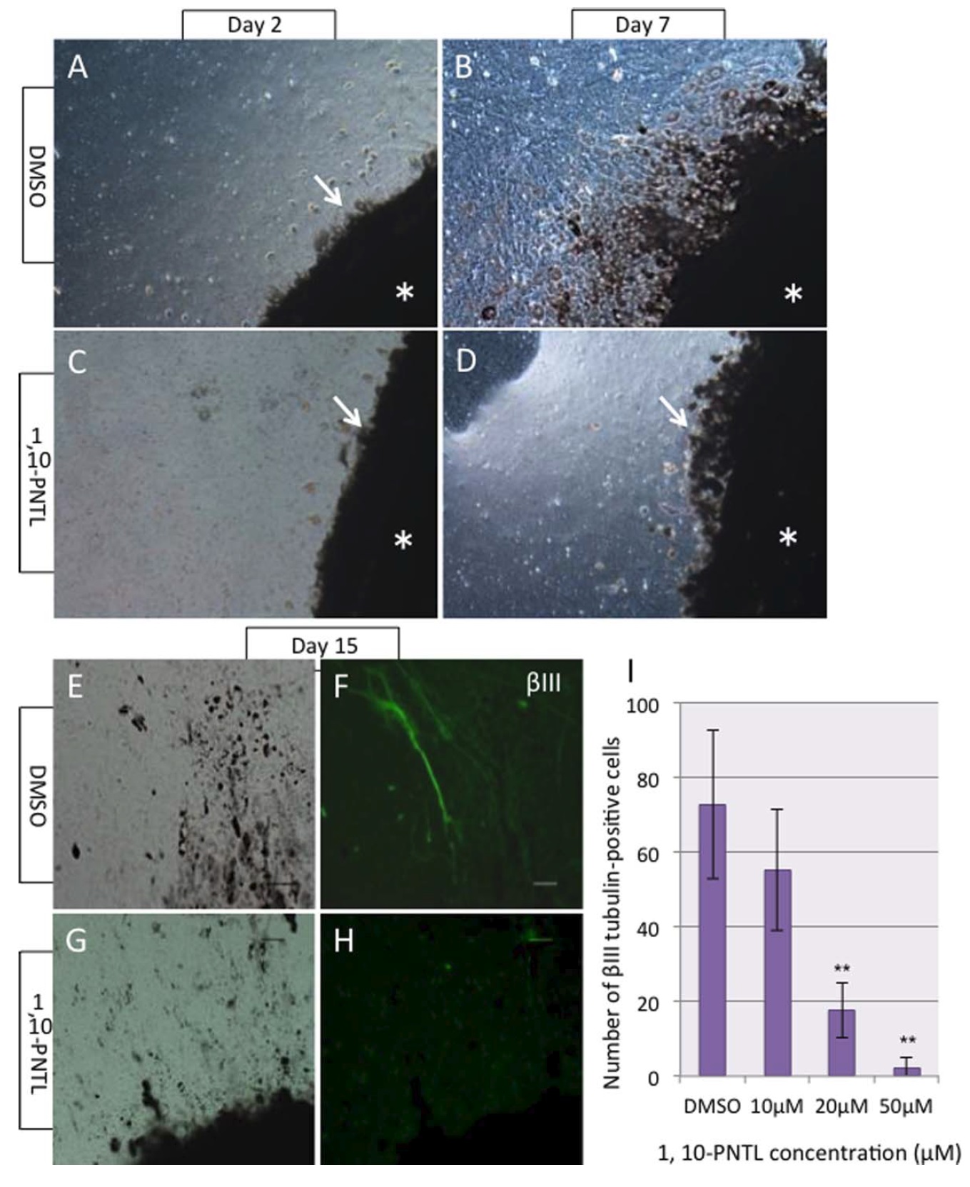
Figure 3 Effects of MMP inhibitor, 1, 10-PNTL on RPE cell migration and neuronal differentiation.
(A, B) In the control culture, RPE cells already began to migrate from the explants on Day 2
(arrow in A) and form the epithelial zone on Day 7. (C, D) In the presence of the inhibitor, only a
few RPE cells were observed outside of the explant (arrows). Asterisks indicate tissue explants. (Eâ
H) Neuronal differentiation was detected by bIII tubulin immunocytochemistry on Day 15. The
inhibitor substantially suppressed differentiation of bIII tubulin-positive cells at 20 mM. (I) Cell
counting of bIII tubulin-positive cells. The data are the mean cell number from 4 explants at each
concentration of the inhibitor. Three separate experiments were performed and the representative
data are shown in the figures. *, p<0.05 vs. cultured without the inhibitor (DMSO). [Color figure
can be viewed at wileyonlinelibrary.com] Image published in: Naitoh H et al. (2017) Copyright © 2017. Image reproduced with permission of the Publisher, John Wiley & Sons. Larger Image Printer Friendly View |
