XB-IMG-128654
Xenbase Image ID: 128654
|
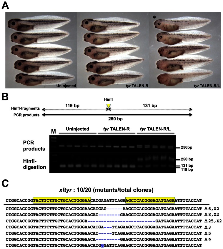
Fig. 1. Disruption of tyrosinase (tyr) in TALEN mRNA-injected embryos.(A) Phenotypes of tyr TALEN-injected embryos: Uninjected control embryos; TALEN-R, embryos injected with 1100 pg right tyr TALEN mRNA; TALEN-R/L, embryos injected with 550 pg of each right and left tyr TALEN mRNA. Injected embryos were reared to the hatching stage. (B) Detection of tyr mutations by genomic PCR and digestion with a restriction enzyme. Five embryos each were collected from the three experimental groups (Uninjected, TALEN-R, and TALEN-R/L). Upper image: a schematic drawing of tyr genomic PCR product. Genomic DNA of each embryo was prepared and then was subjected to PCR with a specific primer set for amplification containing the tyr TALEN target site. HinfI site is located on spacer sequence of the TALEN site. Middle image: a gel electrophoresis image of tyr genomic PCR products. Lower image: HinfI-digestion of PCR products for detecting tyr mutations. PCR products were purified and then were digested by HinfI. HinfI fragments are divided into 131 and 119 bp. M means 100 bp ladder marker. (C) Sequences and frequencies of tyr mutations. PCR product from the TALEN-R/L-injected embryo (asterisk in panel A) was subcloned and was subjected to DNA sequencing analysis. Mutant sequences were aligned to that of wild type. Sequence of wild-type allele is shown at the top. Yellow boxes indicate tyr TALEN target site. Gaps generated by deletion are shown as blue dashes. A substitution is shown as an underlined blue character. Types and frequencies of each mutation (deletion and insertion) are shown at the right of the panel. Image published in: Suzuki KT et al. (2013) © 2013. Creative Commons Attribution-NonCommercial-ShareAlike license Larger Image Printer Friendly View |