XB-IMG-116682
Xenbase Image ID: 116682

|
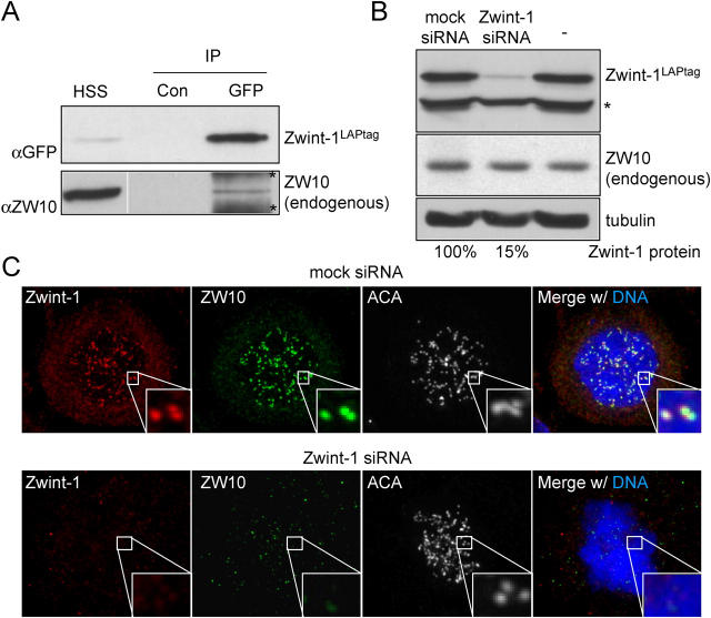
Figure 2. Interaction between Zwint-1 and ZW10 controls ZW10 kinetochore localization. (A) Immunoblot of Zwint-1 immunoprecipitates shows weak interaction with ZW10. Cells of clone LINT2.8 were subjected to immunoprecipitation with control antibody (Con) or anti-GFP antibody to precipitate Zwint-1LAPtag and the precipitate was analyzed for the presence of endogenous ZW10. HSS, high speed supernatant before the immunoprecipitation. Bands labeled with asterisks are background due to precipitation from HSS with the anti-GFP antibody. White line indicates that intervening lanes have been spliced out. (B) Analysis of Zwint-1 knockdown efficiency by immunoblot using cells expressing Zwint-1LAPtag. Lysates of LINT2.8 cells untransfected or transfected with mock or Zwint-1 siRNA plasmid for 72 h were analyzed for Zwint-1LAPtag (anti-GFP), ZW10, and tubulin expression. Percentage of remaining protein was determined by serial dilution immunoblotting. Band labeled with asterisks is protein that cross reacts with anti-GFP in the LINT2.8 cell line. (C) Immunolocalization of ZW10 in cells depleted of endogenous Zwint-1. HeLa cells transfected as in B were treated with nocodazole for 30 min before fixation and stained for endogenous Zwint-1 and ZW10, and for centromeres (ACA) and DNA (DAPI). Image published in: Kops GJ et al. (2005) Copyright © 2005, The Rockefeller University Press. Creative Commons Attribution-NonCommercial-ShareAlike license Larger Image Printer Friendly View |
