XB-IMG-124947
Xenbase Image ID: 124947
|
|
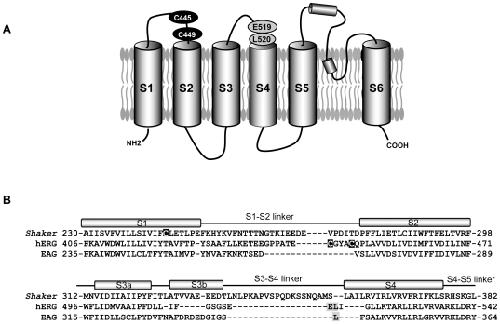
Figure 1. Topology of the hERG channel and location of endogenous cysteine residues.(A) Proposed membrane topology of the hERG channel showing relative positions of endogenous cysteine residues in S1–S2 linker (C445 and C449), and location of introduced cysteines (E519, L520C). (B) Alignment of primary sequences of amino acids of hERG, Shaker and EAG, in the S1–S2 and the S3–S4 regions. The exact positions of the helices and linkers are from [44]. The endogenous cysteine residue removed in Shaker fluorimetry studies (C245) and the accessible cysteine residues in the hERG channel are in white font on dark background. Black letters in gray background indicate sites of introduced cysteine residues and TMRM binding in the hERG (this study) and EAG channels [5]. Image published in: Es-Salah-Lamoureux Z et al. (2010) Es-Salah-Lamoureux et al. Creative Commons Attribution license Larger Image Printer Friendly View |