XB-IMG-127092
Xenbase Image ID: 127092
|
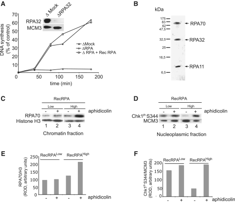
Figure 2. Activation of the replication checkpoint with limited amounts of a recombinant RPA complex. (A, inset) Western blot of egg supernatants after depletion with control (ΔMock) or RPA-specific antibodies (ΔRPA32). (A) Kinetics of DNA synthesis of egg extracts depleted with either control antibodies (ΔMock), or RPA antibodies (ΔRPA) reconstituted with a recombinant RPA complex (ΔRPA + Rec RPA). (B) Silver stain of the RPA recombinant complex. Arrows indicate the three RPA subunits. (C) Western blot of chromatin fraction obtained upon incubation of sperm chromatin in egg extracts depleted with RPA antibodies (A) in the absence (−) or presence (+) of aphidicolin and reconstituted with low or high amounts of recombinant RPA complex (Rec RPA). (D) Analysis of Chk1 phosphorylation in nuclear soluble fractions of the experiment described in panel (C). (E) Quantification of the level of RPA accumulation onto chromatin of the experiment described in (C). Western blot signals were quantified by densitometry scanning and expressed as relative optical density (ROD) compared to the histone H3 signal as loading control. (F) Quantification of the level of Chk1-PS344 in nuclear soluble fractions of the experiment described in (D). Western blot signals were quantified and expressed as relative optical density (ROD) compared to the MCM3 signal that serves here as loading control. Image published in: Recolin B et al. (2012) © The Author(s) 2011. Creative Commons Attribution-NonCommercial license Larger Image Printer Friendly View |