XB-IMG-126030
Xenbase Image ID: 126030
|
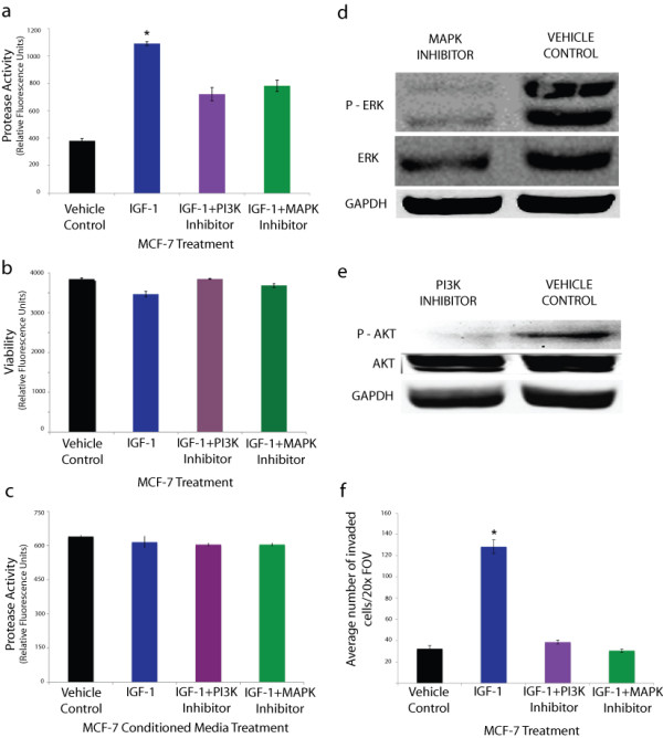
Figure 1. IGF-1 increased metalloproteinase activity and invasiveness in MCF-7 breast cancer cells via the PI3K and MAPK pathways. a) Treatment of MCF-7 cells with 100 nM recombinant IGF-1 caused a 2.9 fold increase in protease activity as determined using a fluoregenic metalloproteinase substrate. Pretreatment of cells with PI3K or MAPK inhibitors attenuated the increased protease activation and resulted in a 34% and 29% decrease in activity respectively compared to IGF-1 treatment alone. b) MCF-7 cell viability, assessed by a fluorogenic viability assay, was not significantly affected by IGF-1 and/or inhibitor treatments. c) Treatment of MCF-7 conditioned media with aforementioned reagents resulted in no significant changes in MMP activity. d-e) Western Blot analysis of MCF-7 cells treated with MAPK or PI3K inhibitors reveals decreased expression of phospho-ERK and phospho-AKT expression respectively. f) Treatment of MCF-7 cells with 100 nM recombinant IGF-1 increased invasiveness through a matrigel coated transwell chamber by ~400% compared to vehicle treatment (control). Pre-treatment of cells with PI3K or MAPK inhibitors before IGF-1 treatment resulted in no significant change in invasiveness compared to vehicle control. Each assay was repeated three times (three experimental repeats). All data are mean ± s.e.m, (n = 3) *P < 0.01. Image published in: Walsh LA and Damjanovski S (2011) Copyright ©2011 Walsh and Damjanovski; licensee BioMed Central Ltd. Creative Commons Attribution license Larger Image Printer Friendly View |