XB-IMG-122952
Xenbase Image ID: 122952
|
|
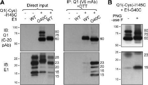
Figure 5. (A) Confirming the presence of E1 G40C in the 80- and 75-kD Q1-positive bands detected in whole cell lysate of COS-7 cells coexpressing Q1(-Cys)-I145C and E1-G40C. COS-7 cells are transfected with the cDNA(s) listed on top. An aliquot of the whole cell lysates is loaded onto the “direct input” lanes. The other aliquot is subjected to immunoprecipitation with V5 mAb. The immunoprecipitates are DTT treated and loaded onto the IP lanes. The Q1 and E1 are probed using a goat pAb against Q1 (C-20) and a rabbit pAb against E1. (B) Testing N-glycosylation in free E1 proteins and disulfide-linked Q1/E1 complex. Whole cell lysates from COS-7 cells coexpressing Q1(-Cys)-I145C and E1-G40C are treated with PNGase F or with buffer under the same conditions (“+” and “−” PNGase F) for 48 h at 37°C. The samples are fractionated by nonreducing SDS-PAGE and probed for Q1 and E1. Image published in: Xu X et al. (2008) © 2008 Xu et al. Creative Commons Attribution-NonCommercial-ShareAlike license Larger Image Printer Friendly View |